
Prof. Dr. med. Dr. h.c. G. Ulrich Exner
FMH Orthopädische Chirurgie
und Traumatologie
Minimally invasive techniques
The development of cross sectional imaging techniques (especially the development of Computed Tomography – CT Scan) has greatly expanded the navigational armamentarium. In the field of Tumor Orthopaedics the use of Computed tomography has been contributed that open biopsy in moste cases could be replaced by needle biopsies. The great advantages of needle biopsies over open biopsies are not only the minimizing of contamination but even more the precise orientation in taking the specimen and documentation where the material was taken.
Minimally invasive techniques can also be applied in the treatment of certain lesions . Professor Exner has greatly contributed to the introduction of computed tomography (CT) guided drill excision and radiofrequency ablation of osteoid osteomas.
Radiofrequency (RF) treatment is also of increasing importance in the treatment of metastases.
Bone tumors (e.g. malignant tumors as osteosarcoma, EWING sarcoma, and chondrosarcoma, or aggressive benign tumors as giant cell tumors, aneurysmal bone cysts, vascular malformations)
In malignant tumors it is mandatory that no tumor is left even macroscopically (R0 resection = residual tumor yero). This means, that large segments need be removed with a healthy tissue layer around the tumor without opening or touching the tumor itself.
Professor Exner has made important contributions to the development of loca resections and reconstructions preserving the extremities in place of amputations needed formerly.
The easiest is to replace large defects with massive endoprostheses. However these are subject of wear and loosening. Therefore only biologic reconstructions can lead to permanent solutions.
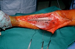
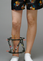
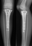
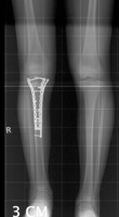
This 6 year old boy suffered from an osteosarcoma of the tibia. He was treated by local resection, reconstruction with an allograft covered by a gastrocnemius flap to protect the allograft and reconstruct the extensor mechanism.
Because of the loss of the growth plate shortening of the leg developed which was treated by lengthening of the affected leg and contralateral epiphysiodesis (minimally invasive percutaneous growth plate destruction).
12 years after treatment the boy is disease free has equal leg length and has no physical limitations.
Osteosarcoma of the tibia
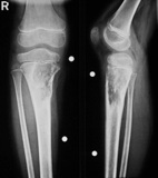
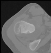
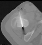
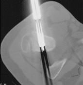
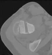
Osteoid osteoma of the posterior aspect of the femoral neck.
This 8 year old gurl suffered from an osteoid osteoma of the posterior aspect of the femoral neck. These benign tumors – if they are in the vicinity of joints – can cause joint degenation similar to the mechanisms as rheumatoid arthritis.
Open resection usually needs a very large exposure and – because they are very small – frequently are missed at open surgery. They are excellently depicted by CT and it is logic to use this ‚navigation’ vor the removal. The safest way is of course chosen which may not always bet he shortest as in the patient shown here.
The most important principle always is to keep the damage as small as possible. Whenever applicable minimally invasive techniques including the necessary navigation are chosen as is also done fort he artificial joint replacement.